Equipment

Molecular Histology and Fluorescence Imaging Equipment
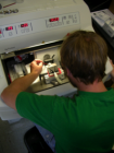
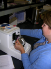
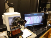
The MHFI core provides the resources and expertise for preparing tissue for histological staining. We offer routine fixation, embedding, sectioning and staining of tissue specimens- either paraffin embedded or frozen tissue. The Imaging component provides researchers with analyzable images of cells/ tissues taken from both the light and confocal microscopes. All of the microscopes in MHFI are interfaced with computers with proprietary software for collecting digital images. We also have copies of the software installed on workstation computers for post-collection analysis.
The instruments present in Histology include:
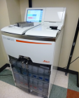
- Leica ASP 300 Tissue Processor
- Leica CM1950 cryostat
- Leica RM2235 & Shandon Finesse 325 rotary microtomes
- Leica ST5010 Autostainer
The core employs a staff scientist versed in histology and immunohistochemistry (IHC) techniques. The scientist consults with researchers on experimental design, maintains the core equipment, provides histology services and training to researchers and students, and other related services as needed. Equipment use/ sign-up sheets are used in Histology.
Core Imaging instruments include:
- Nikon E 800 epi-fluorescent/transmitted light microscope, with an Olympus DP71 camera with cellSens software
- Nikon TE 300 inverted microscope with DIC and epi-fluorescence
- Olympus FV 1000 IX inverted laser scanning confocal microscope with 405, 458,488,515, 559 & 635 nm laser lines
- Olympus SZX16 fluorescence dissecting microscope, with Olympus DP26 camera, cellSens software
- CytoViva hyperspectral microscopy and imaging system
- Software available on three workstation computers includes Image J, Photoshop, Triple I Slidebook and Image Pro Plus V7.0
The facility is operated under the direction of Dr. Scott Wetzel with Lou Herritt (Staff Scientist) providing technical assistance.
The MHFI Core is located in the Skaggs Building - rooms 060C, 070 and 052A.


