
Hyperspectral Imaging
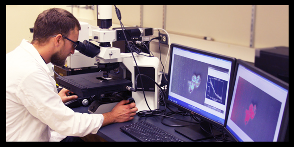
Providing hyperspectral microscopy and imaging capabilities to researchers, the CytoViva includes a darkfield-based light illumination system, epi-fluorescence with three colors (blue, green and red) that can be combined and a hyperspectral microscope. Spectral image libraries can be used to characterize sample nanoparticles, pathogens or subcellular materials.
The CytoViva can image objects down to 10 nm in size and the corresponding image analysis software enables mapping the sample elements based upon their unique spectral characteristics. Hyperspectral microscopy can then be used to determine the location of nanoscale materials within various biological samples (see image gallery below).
For more information on the possibilities, visit: http://www.cytoviva.com
If you are interested in utilizing the CytoViva in your research, please email CEHS Biostatistician Ray Hamilton or Staff Scientist Lou Herritt.
Funding for the CytoViva system was made possible with a grant from the M. J. Murdock Charitable Trust.
Image Gallery


Main menu
![]()
